|
|
||||||||||||||
|
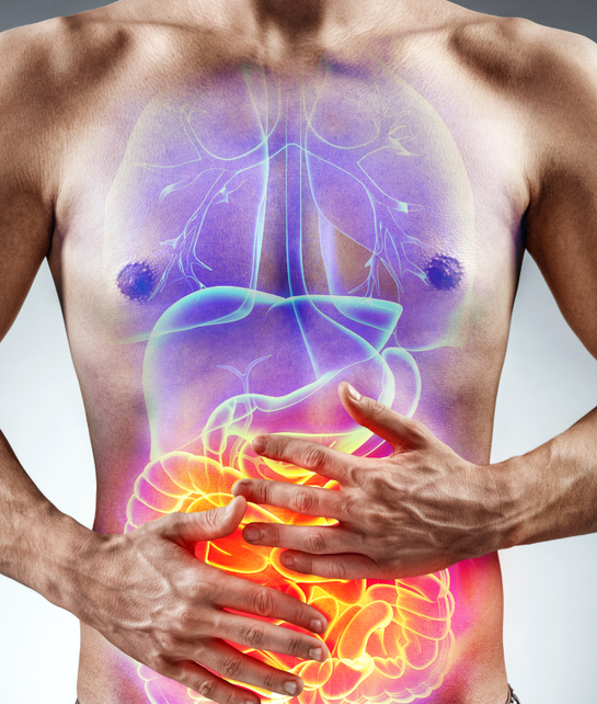
Types of bladder cancer |
Home Treatments Lifestyle Symptoms Cancers | |||||||||||||
This refers to what it looks like down a microscope and includes the histological type and the histological grade. These factors in combination with how advanced the tumour has grown (stage), the general condition of the patient, determine the prognosis (likely outcome) and the best modality of treatment for that individual. Histological type - There are three types of bladder cancer that start in the lining of the bladder depending on the cells that become cancerous:
Histological grade - The pathologist will determine the aggressiveness or grade of the tumour by comparing the microscopic features of the surgical specimen with an established classification system. In general these systems look at: How the pattern of cells within the tumour looks the organ of origin - if a low grade (often otherwise called grade 1, or well differentiated) tumour will have features, albeit disrupted, of it origin. If high grade (grade 3 or poorly differentiated) will just look like a sheet of cells with no residual distinctive pattern remaining. How aggressive the cancer cells look like. If the cells are large, show features of dividing rapidly or are very dissimilar to those from its organ of origin they are usually classed as high grade. If small, look like they are they are not dividing rapidly and look similar to those from its origin they are usually be classed as low grade. Often more advanced test are performed at this stage by the pathologist using special stains or process picking up receptors on the cell surface or nucleus called immunohistochemistry. A good example of these or oestrogen or HER2 receptors for breast cancer but similar less known immunohistochemical tests can be used for a wide range of cancers. Further general information Your doctors and specialist nurses are in an ideal position to give you relevant information on your disease and treatment as they know your individual circumstances. Cancerbackup has a help line (0808 800 1234) and a prize winning video available in English, Italian, Urdu, Bengali, Gujarati & Hindi explaining Radiotherapy & Chemotherapy. Cancernet.co.uk has over 500 pages describing cancer, its management, practical tips and tool which patients, their carers and their doctors have found helpful during the cancer journey. |
||||||||||||||