|
|
||||||||||||||
|
Advanced radiotherapy techniques |
|
|||||||||||||
| Home | Cancer treatments | Lifestyle | Site map | Contact us |
|
|||||||||||||
| Contents: Conformal radiotherapy | Tomotherapy | IMRT | IGRT | Prostate brachytherapy | ||||||||||||||
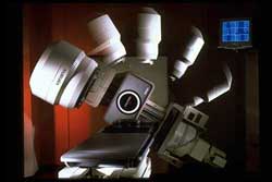
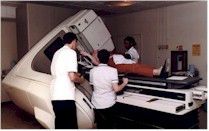
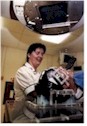
The majority of radiotherapy treatments originate from machines called linear accelerators. These are large and sophisticated which produce high energy x-rays (or photons). The are also expensive and require a lot of highly trained manpower to keep them working accurately. The head of the machine, bed and gantry can be rotated and moved in multiple directions to ensure the radiotherapy beam can enter the body in virtually any direction. In the passed the beam only came out in square shapes. Conformal
radiotherapy. This is now routine technology available in the vast
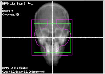
majority of radiotherapy centres throughout the world. Lead blocks or This is an example of a patient with cancer of the air sinuses of the face. The green line is shape of the beam coming out of the linear accelerator but the purple line is eventual shape after it has been modified by the lead blocks in order to conform the shape of the target. In this case, this has avoided irradiating the eyes which may course cataract and dryness later in life and the salivary glands causing an uncomfortable dry mouth. Virtually all linear accelerators in clinical practice are now capable of conformal radiotherapy. Not all machines are capable of two further recent scientific developments: Intensity modulated radiotherapy (IMRT) The next generation of machines are even more accurate, capable of varying the intensity of the beam within the area treated area. In this way, the contours of the body are taken into account avoiding hot and cold spots within the treated field. This is not optimal as the hot spots could damage the normal skin causing fibrosis and pain. On the the other hand the cold spots could not kill the cancer cells in this area allowing it to not be locally controlled and increase the risk of it returning after treatment or spreading to another part of the body. Tomotherapy At the time of writing this page (Feb 2009) there are only two tomotherapy machine sin the UK (Addenbrooke's Hospital Cambridge and the Cromwell hospital, London). These are the most sophisticated machines commercially available They are capable of changing the both the shape and intensity of the beam during the actual treatment. As a result they can produce a very accurate distribution of radiotherapy in the body. In stead of 2-4 beams it tend to use multiple constantly changing beams. This machine is enabling doctors to treat tumours which previously were not possible due to their proximity to other vital organ which can now be protected. They also have more accurate image guidance systems. Image guided radiotherapy (IGRT). Normally the radiotherapy beam is "set up" to the correct position using tattoos on the skin or marks on a shell. Although generally accurate, errors can be made if the skin is loose or moves due to breathing or changes in shape of the patient. Sometime the target organ or cancer can move during the treatment. A good example of this is the bladder depending how much urine it contains or the prostate which can move depending how much gas or feaces there is in the rectum. IGRT uses a technique which guides the radiotherapy using a target within the cancer or organ itself so it doesn't matter so much if it has moved between days of treatments. Several techniques are used for this including CT imaging on the linear accerator itself or the insertion of a gold grain for example in the prostate before treatment. |
||||||||||||||